


New plastic surgery simulation and imaging technologies are allowing medical professionals to prepare for complex procedures like never before, while giving patients a better sense of the expected surgical outcomes.
It’s not easy to imagine what we’ll look like with a new nose, chin, or chest. Sure, we can spy that coveted look on someone else or picture it in our mind’s eye, but the reality doesn’t always live up to our expectations.
Computer scientists and medical professionals are continuing to perfect 3D imaging, augmented reality, and other related technologies in the effort to not only provide patients with more accurate projected results of their upcoming surgeries, but also to improve the surgical techniques themselves.
Revolutionizing the Consultation Process
Imagine visiting a surgical center for a rhinoplasty consultation. You and the doctor are discussing the changes that you want to make. But instead of pulling out a collection of photographs of recent successful surgeries or showing you 3D printed models of the various popular approaches, your doctor leads you to a machine that looks like it belongs on board the Starship Enterprise.
He or she then directs you to stand facing a series of screens. The machine snaps your photo from several angles and combines them into one three-dimensional image. Soon you’re consulting with the doctor, staring at a rotating digital version of your own head.
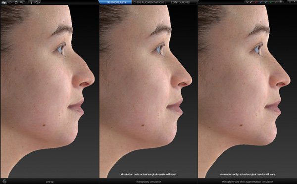
The likeness is uncanny, right down to the freckles on your cheeks. The doctor changes the 3D image to show you how you’ll look with various noses. You no longer need to rely on your imagination.
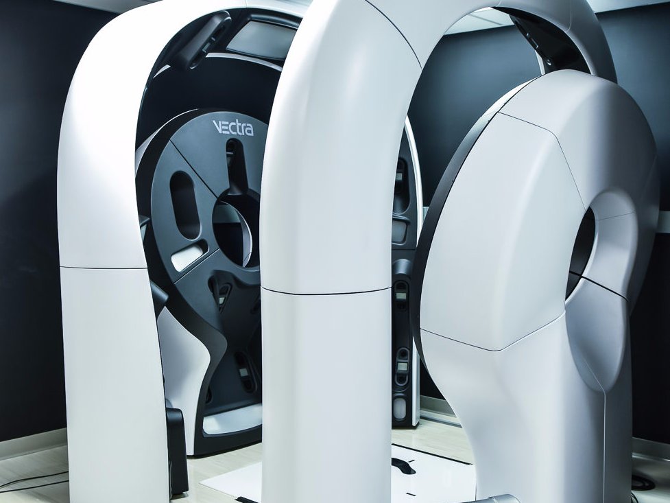
This is the Vectra 3D Imaging System. When Canfield Scientific Inc. first launched the technology in 2009, it could only simulate breast augmentations. However, it has since expanded to include breast lifts, rhinoplasties, chin augmentations, and even liposuction.
By taking six two-dimensional photographs of your face and body, the Vectra 3D Imaging System can recreate your entire body in digital 3D with remarkable precision.
Although the technology is primarily intended for use during consultations, it also has benefits for the surgeries themselves. Noting that it’s sometimes difficult to detect asymmetries with the naked eye, some doctors have credited 3D imaging with making surgery more precise and efficient.
In short, it helps both doctors and patients see the desired outcomes of their surgeries far more accurately.
Smartphone Apps: You Pay for What You Get
Of course, state of the art technology doesn’t always come cheap. One 2014 study sought to determine if Vectra 3D Imaging Systems were more accurate that less expensive alternative. Researchers compared three-dimensional digital renderings of a mannequin made by Vectra with those of Autodesk 123d Catch, a smartphone app that creates 3D renderings from smartphone photos.
The study deemed Vectra faster and more efficient. What’s more, the Autodesk 123d Catch lacked the tools necessary to effectively manipulate the image, as would be required in a consultation.
Nevertheless, that a three-dimensional image could be rendered using only a smartphone is a testament to how far this technology has evolved over the course of a few short years. “Soon mobile applications may offer an alternative for plastic surgeons to today’s cost intensive, stationary 3D camera systems,” researchers concluded.
The Autodesk 123d Catch was ultimately discontinued in January 2017, but the technology persists and AR Systems, the company responsible for the app, has plans to repurpose the technology in future apps. It’s not impossible this technology will soon be accessible enough to become a staple of every plastic surgery facility.
Soon mobile applications may offer an alternative for plastic surgeons to today’s cost intensive, stationary 3D camera systems.
Critics of the Technology
While many swear by the technology, some studies suggest that 3D imaging has no appreciable effect on patient satisfaction for certain plastic surgery procedures. This 2015 study in Plastic and Reconstructive Surgery looked at breast augmentation patients who consulted doctors with and without this technology.
The researchers concluded that patient satisfaction actually declined by approximately 25 percent among those who received consultations with 3D imaging.
These researchers concluded that the three-dimensional images failed to accurately depict real-life surgical results. They pointed out that different tissue can react differently to the same breast implants, something 3D imaging technology would not be able to account for. It’s also possible that patients were holding the surgery to a more specific, higher standard in light of the virtual preview they saw.
In either case, this doesn’t change the fact that for the time being 3D imaging provides patients with the most accurate advance view of the results of their respective surgeries. Neither does it change any of the other remarkable benefits 3D imaging brings to the surgical table.
The Expanding Use of 3D Imaging in Medicine
Three-dimensional imaging allows surgeons to see and manipulate the human body in a safe, digital environment. The potential for this technology goes far beyond setting realistic expectations for patient outcomes. Recently, three-dimensional imaging has been used to support some highly complex and risky procedures.
Earlier this year, surgeons at the All India Institute of Medical Sciences (AIMS) in New Delhi used this technology to separate conjoined twins Honey and Singh Kanhar (pictured below). This unusual case involved infant twins that were joined at the head – a rare condition. When treated, however, the relatively dismal rate of success for this type of surgery currently falls below 25 percent.
The surgeons involved in this particular instance studied three-dimensional images created from CT scans, also known as CAT scans, MRI scans, and angiography X-ray imaging. These images allowed the surgeons to see inside of the twins’ heads to better prepare for the surgery. They were even able to don HTC Vive virtual reality (VR) headsets and Haptic robotic arms to practice the surgery. The technology simulated the feel of actual surgery to promote a more immersive experience.
The first stage of the surgery, which involved separating the brains and venous channels, was a complete success. The second stage, separating the skulls completely, had yet to be performed at the time of this writing, but the prospects are promising.
A similar, albeit less risky, surgery was performed in Corpus Christi, Texas, in 2016. Conjoined twins joined at the hip were successfully separated thanks in part to 3D Systems. CT scans of the twins were translated into a three-dimensional digital rendering. Using these scans the surgeons were able to simulate the procedure and even print anatomical models they could practice on in advance of the operation.
From consultations to risky surgeries, three-dimensional imaging makes it possible to better predict a new reality before putting anyone under the knife. As the technology progresses, it could hold significance for a wide variety of medical procedures – plastic, reconstructive, or otherwise. The technology is progressing rapidly, and perhaps best of all, in ways that promise huge improvements on the plastic and cosmetic surgery stage.

The Future of 3D Imaging with Augmented Reality
You’ve heard of virtual reality, but you may not yet be familiar with the concept of augmented reality. While virtual reality sucks you into a completely virtual world, augmented reality instead enhances the real one with virtual elements. This technology is popular in gaming apps like Pokémon Go. However, it’s also a handy addition for plastic surgeons.
Let’s revisit that rhinoplasty consultation. You and your doctor settle on the perfect nose. It’s time to take the plunge. Your doctor takes the three-dimensional simulation from your consultation and projects it onto your features as you lie on the operating table. Your new nose is overlaying your current one like a hologram. Your doctor uses this as a reference point during the procedure.
Before you get too excited, however, remember that this technology is still in the budding stages. According to media reports, a research group out of Osaka Medical College in Japan has recently tested it. Using high-definition cameras, they generated a three-dimensional image of the surface features of a test subject. They combined this with CT scans for a more accurate facial structure. After they finished rendering a simulation of the ideal results, the researchers projected those results onto the subject, viewing them through a pair of smart glasses.
The technique is said to dramatically improve surgical precision. Based on their observations, augmented reality proved a useful tool for planning and executing a surgery, as well as evaluating surgical results. Of course the researchers’ hopes for the tech don’t stop there. They envision improved surgical training methods and easier ways to measure surgical results.
It’s hard to imagine that any technology with such wide-reaching applications and dramatic benefits wouldn’t be around for the long haul. And you can rest assured that while the technologies in question are still largely in the embryonic stage, soon, from practice and planning to execution, 3D imaging will revolutionize the entire surgical process.